
Principais cirurgias da coluna cervical Dr. Alberto Gotfryd
C5 6 Stock Photos and Images (707) See c5 6 stock video clips Quick filters: Cut Outs | Vectors | Black & white c4 5 c5 6 anterior cervical discectomy c3 4 c5 6 and c6 c7 disc herniations Sort by Relevant
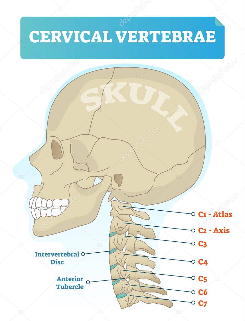
Vértebras cervicales vector ilustración. Esquema con cráneo, atlas C1
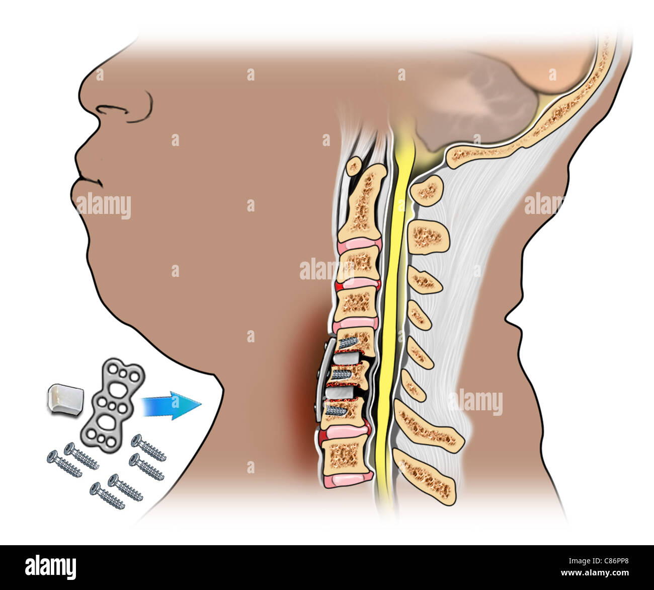
The fifth and sixth cervical vertebra (C5-C6) is the most easily injured segment encountered in clinical practice. The anterior cervical plate and cage (ACPC) fixation system is always used to reconstruct the intervertebral height and maintain the segmental stability. The postoperative effect, such as subsidence, neck pain, and non-fusion, is.

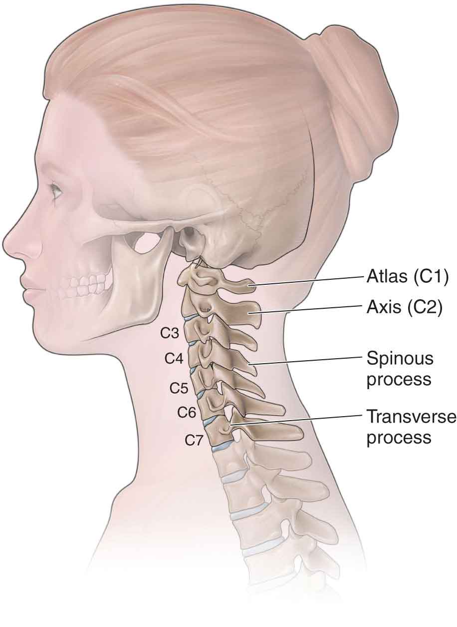
C1 a C7 A coluna cervical é composta por 7 vértebras sequenciais, que
These spinal bones attach to the thoracic spine and work together to support the head. The sixth cervical vertebra or C6 is relatively smaller in size (compared to remainder of cervical vertebrae.

Cervical Xray Showing Spinal Fusion C5 C6 C7 Foto de stock Getty Images
See C5-C6 Treatment. C6-C7 (C7 nerve root): Pain, tingling, and/or numbness may radiate into the hand and middle finger. Weakness may also be felt in the triceps (muscles in the back of the upper arm), finger extensors, and other muscles. The C6-C7 disc is commonly considered the most likely to herniate in the cervical spine. 1 Rainville J, et.
muscles of cervical spine
Cervical nerves are spinal nerves that arise from the cervical region of the spinal cord. These nerves conduct motor and sensory information via efferent and afferent fibers, respectively, to and from the central nervous system. While classified as peripheral nerves, the motor cell body resides in the anterior horn of the spinal cord. There are eight pairs of cervical nerves, denoted C1 to C8.

Cervical vertebrae C6 stock photo. Image of anatomy, spine 81703964
Click To View Large Image. The C6 vertebra is the sixth cervical vertebra of the spine. It is found in the base of the neck between the C5 and the last cervical vertebra, C7. The C6 vertebra plays an important role in supporting and protecting the structures of the head and neck as well as anchoring the muscles that move and support the neck.

Cirurgias na coluna cervical Colunar
Cão da raça lhasa apso com 2 anos de idade apresentando quadro severo de compressão cervical. Após localização exata da lesão pela tomografia computadorizada.

Xray anteroposterior and lateral views of cervical spine showing
The C6 myotome is a group of muscles controlled by the C6 nerve. These muscles include the wrist extensor muscles, which allow the wrist to bend backward; and the biceps and supinator muscles of the upper arm, which serve to bend the elbow and rotate the forearm. See Cervical Spinal Nerves
Anatomía de las cervicales. Conoce sus huesos, ligamentos
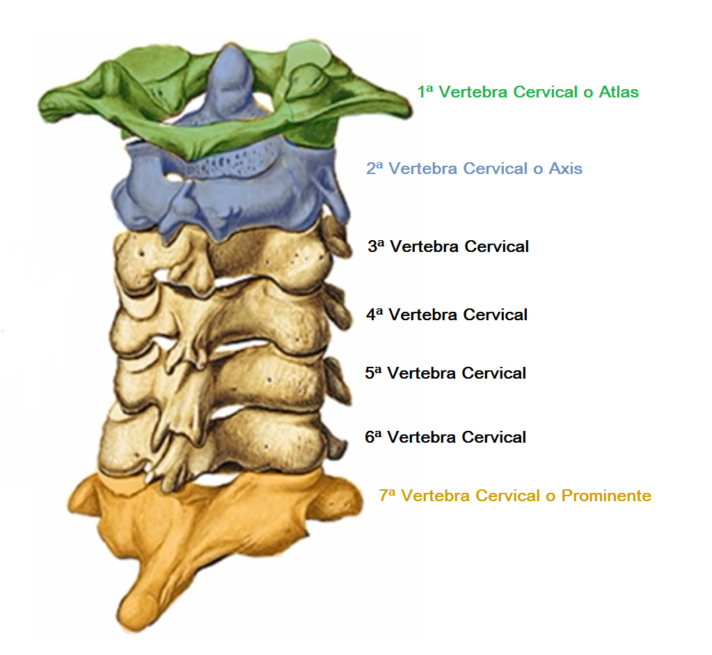
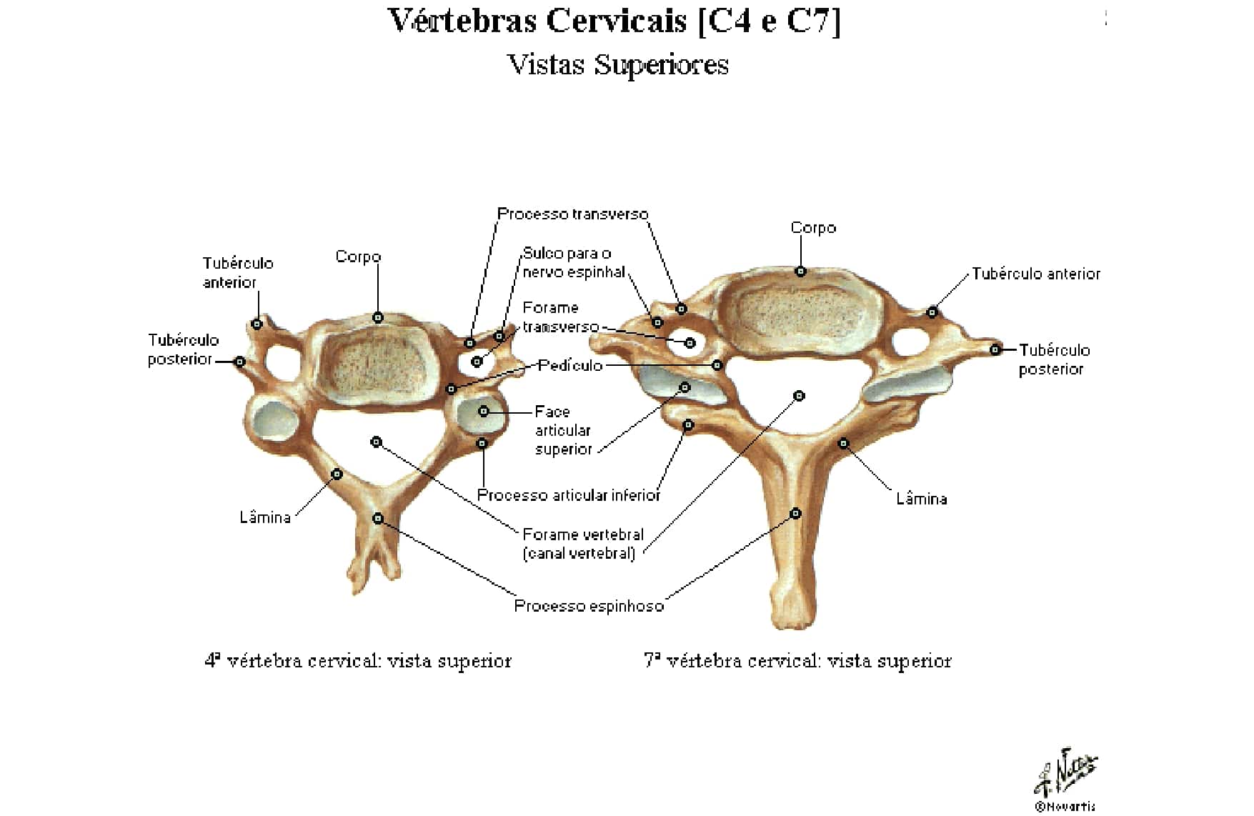
Cervical vertebrae Typical cervical vertebrae (C3-C5) Atlas (C1) Axis (C2) Sixth cervical vertebra (C6) Vertebra prominens (C7) Thoracic vertebrae Typical thoracic vertebrae (T2-T9) Atypical thoracic vertebra: T1 Atypical thoracic vertebra: T10
Cervical Bone Fusion of C56 and C67 Stock Photo, Royalty Free Image
A pinched nerve on C5-C6 that roots in the neck impacts the neck, arms, hands and shoulders with radiating pain. Often, tingling and numbness in these areas are another common symptom of C5-C6 disc bulging. This can lead to muscle weakness and spasms and additional discomfort in the digits of the hands. If you are experiencing any of the.
Vértebras cervicais Anatomia papel e
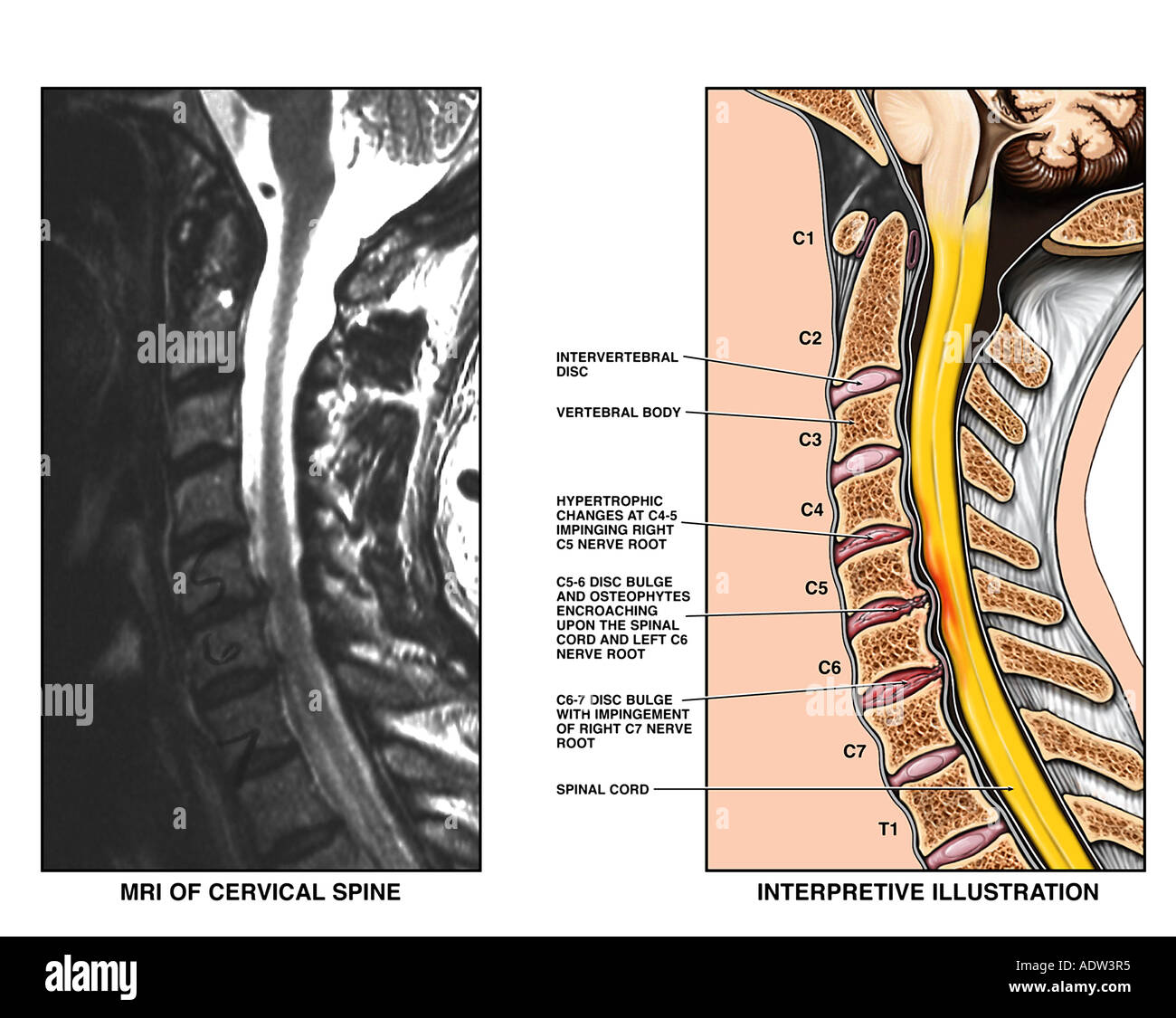
When a cervical disc herniates, the main symptom is localized or radiating pain. However, each vertebral segment can be characterized by specific symptoms. In the case of herniation of the C5-C6 disc, the symptoms are as follows: Weakness in the biceps. Pain, numbness and tingling that runs down the arm, to the forearm, even to the thumb and.
Cervical disc herniations c4 c5 c6 c7 hires stock photography and
C5, C6 medial branch. The base of the neck is scanned in the transverse plane and the TP of T1 identified. As the probe is moved cephalad from this point, the TP of C7 is localized, followed by the targets on the AP of C6 and C5.. Cervical medial branch block: a novel technique using ultrasound guidance. Reg Anesth Pain Med. 2012;37:219-223.

Anatomía de la columna cervical Dolopedia
A coluna cervical consiste em uma das subdivisões da coluna vertebral que, por sua vez, integra o sistema esquelético do corpo humano e desempenha um papel crucial para que todos os membros do corpo se movimentem de forma adequada. Composição da coluna cervical: c1, c2, c3, c41 c5, c6 e c7 A composição da coluna cervical é descrita como:
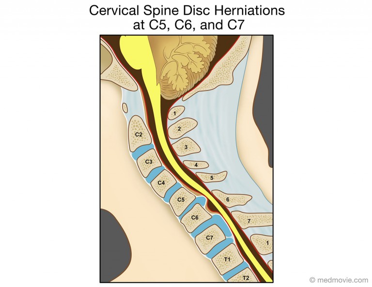
Cervical Spine Disc Herniations at C5, C6, and C7
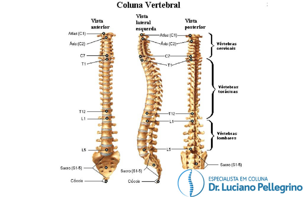
The spine, or vertebral column, is a segmental set of 33 bones and associated soft tissues in the subcranial portion of the axial skeleton. It is subdivided into 5 regions based on curvature and morphology: cervical, thoracic, lumbar, sacral, and coccygeal (see Image. Vertebral Column).
Coluna Cervical Anatomia, Ossos, Ligamentos Kenhub
Foraminal narrowing of the cervical spine. Foraminal narrowing commonly takes place in the C5 to C6 levels of the spine, which is located beneath the middle of the cervical spine and offers structural support and flexibility to the neck. If the foramina become narrowed at this spinal segment, compression can occur on the C6 nerve root, causing.
Dor na Coluna Cervical ou Cervicalgia Causas, sintomas
Pathology. The cervical spine is susceptible to injury because it is highly mobile with relatively small vertebral bodies and supports the head which is both heavy and acts as a lever. C2 (~30%) and C7 (~20%) are the most commonly fractured levels 7. There are many types of cervical spine fracture, some of which are unstable; general indicators.